Cerebellar Mutism
Cerebellar mutism is a disorder that may occur in children being operated for a tumor in the cerebellum. They are fully awake and cooperative but cannot speak. Their unability to communicate is devastating for both the parents and the child, especially during this episode of fear and anxiety. In addition, mutism is frequently followed by severe cognitive and psychological disturbances in the long term.
Language and speech functions have always been attributed to the left cerebral hemisphere. The role of the cerebellum in these functions is still unclear.
The main goal of the research conducted in the Neurosurgical Centre Nijmegen by Kirsten van Baarsen (MD), is to map the cerebellar white matter fibers that are involved in the development of cerebellar mutism. This knowledge will contribute to better diagnosis and treatment, and perhaps even prevention of cerebellar mutism.
In order to compare white matter fibers between subjects, a digital three-dimensional atlas has been developed in collaboration with the Oxford Centre for Functional Magnetic Resonance Imaging of the Brain. It is based on MRI data of 90 subjects participating in the Human Connectome Project.
The atlas is compatible with MNI152 T1 1mm template as well as the SUIT template and it is freely available for researchers in the field. Methodological details will be published shortly.
MNI_1mm_parcellation_map_thrP10.nii
MNI_1mm_parcellation_map_thrP50.nii
MNI_1mm_parcellation_map_thrP90.nii
SUIT_1mm_parcellation_map_thrP10.nii
SUIT_1mm_parcellation_map_thrP50.nii
SUIT_1mm_parcellation_map_thrP90.nii
These atlases are best visualized using ITK snap, click here for download)
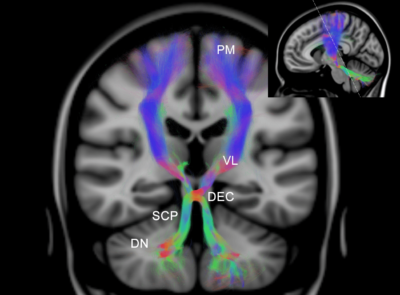
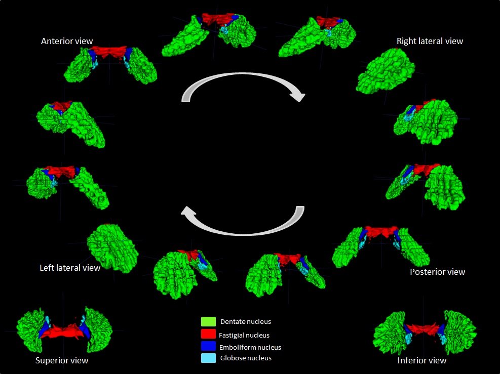
DN, dentate nucleus
SCP, superior cerebellar peduncle
DEC, decussation
VL, ventrolateral nucleus
PM, premotor area
Update march 2016:
Dr. Kirsten van Baarsen: “In our search for the anatomical substrate of the cerebellar mutism syndrome, we came across the lacking of a detailed, digital, three-dimensional atlas of the cerebellar nuclei.
The cerebellar nuclei appear to have a crucial role in the working mechanism of the cerebellum. To understand their function, thorough knowledge of their morphology and anatomical relations is mandatory. Imaging studies of the cerebellar nuclei can be very helpful to appreciate their anatomy but they face several challenges. The nuclei are very small, they have low contrast and their three-dimensional morphology and anatomical relations are complex. Histological studies provide great detail but lack three dimensional information. MRI, on the other hand, does indeed provide this three-dimensional information, but lacks morphological details. Therefore, the correct identification of the individual cerebellar nuclei in MRI remains challenging.
In this work, a three-dimensional histological reconstruction of the cerebellar nuclei was registered with a high resolution MRI of the same post mortem specimen – combining the best of both worlds.
The work has resulted in highly detailed digital three-dimensional atlas, that will be freely available to researchers in the field”.