Innovative, 3-dimensional visualization of neuroanatomical structures: a new app to cover it all!
The anatomy of the brain forms a challenge for many doctors and scientist in their daily practices. Therefore, it is not hard to understand that students struggle with comprehending the anatomy of neural structures. Nevertheless, knowledge of the neuroanatomy is regarded as a hot societal topic as this field of expertise is thought to contribute to our understanding of neurological diseases. When students therefore study the anatomy of the human brain, they use serial sections, depicted in anatomical atlases. The sections are well organized but provide only limited insights in the true, three dimensional anatomy of the human brain.
In order to provide these crucial insights, the departments of Anatomy and Neurosurgery of the Radboud University Medical Center join their forces. PhD-candidate Dylan Henssen and Guido de Jong, technical physician combine classic anatomical knowledge with new technological insights and create a digital, three-dimensional model of the human brain within a smartphone application. This project is empowered by a Proeftuin-grant of the Radboud University.
With this application, students can study the anatomy of neural structures at anytime, anywhere. This virtual reality tool is thought to help them to gain more insights in the three dimensional relations of these structures. Next to this, the department of Anatomy will implement these educational innovations during the standard anatomical courses at the dissection rooms. Finally, an extra-curricular course is scheduled in order to take a group of interested students on a virtual tour through the basal ganglia by using augmented reality.
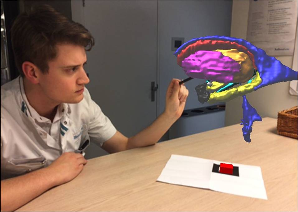
PhD-candidate Dylan Henssen studies the first three-dimensional, digital model of the basal ganglia, limbic system and the ventricular system.